
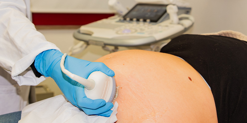
Ultrasound is a safe and non-invasive technique that uses high-frequency sound waves to visualize internal organs and, during pregnancy, the fetus. When the ultrasound waves pass through the tissues, they produce echoes that are transformed into images on the monitor.
The first-trimester scan is performed between 11+0 and 13+6 weeks, generally via the transabdominal route, although in some cases a transvaginal approach may be necessary to obtain clearer images.
This ultrasound allows the verification of fetal vitality, precise dating of the pregnancy, measurement of nuchal translucency, and an initial assessment of fetal anatomy. If the expectant mother chooses to undergo prenatal screening, it can be combined with NIPT (Non-Invasive Prenatal Test), which provides an estimate of the risk of chromosomal abnormalities: trisomy 21 (Down syndrome), 18 (Edwards syndrome), and 13 (Patau syndrome). The third-trimester obstetric ultrasound is performed based on specific indications.