
Morphological Ultrasound

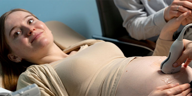
Morphological ultrasound is one of the most important screening tests during pregnancy. It is performed between the 19th and 21st weeks of gestation and aims to assess the vitality, anatomy, and development of the fetus, as well as to check the amount of amniotic fluid and the location of the placenta.

This exam is fundamental because all pregnant women have a general risk of fetal malformation, estimated at about 2–3 pregnancies out of 100. The purpose of the morphological ultrasound is to identify, among apparently healthy fetuses, those at risk of anomalies in order to offer appropriate diagnostic pathways.

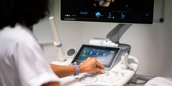
During the examination, several parts of the fetus’s body (such as the head, abdomen, and femur) are measured, and the values are compared with reference curves to verify that the measurements are consistent with the gestational age. The location of placental insertion, the amount of amniotic fluid, and the structure of major organs and anatomical districts are also evaluated.

Screening ultrasound does not allow for the detection of all fetal malformations: only a portion of congenital anomalies are identified prenatally, and some may appear later in pregnancy or even after birth. Factors such as fetal position, reduced amniotic fluid, maternal obesity, or abdominal scars can reduce image quality.


Moreover, the purpose of the morphological ultrasound is not to detect genetic abnormalities, as many of these are not associated with sonographically visible malformations. Although it has a precise clinical purpose, the morphological ultrasound also represents a special moment for the couple. Our goal is to offer an accurate and reassuring service, using state-of-the-art technology and the expertise of a specialized team, in a comfortable and welcoming environment.